CN: 14-561
Species: Equid, Horse
Specimen:
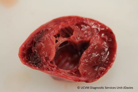
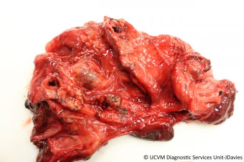
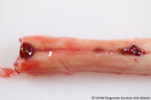
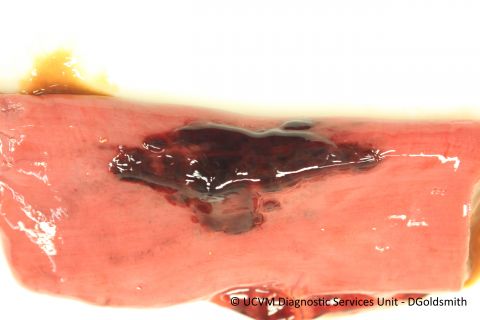
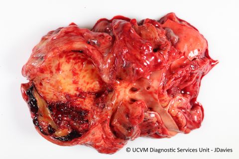
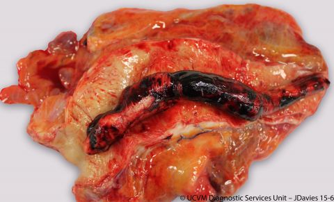
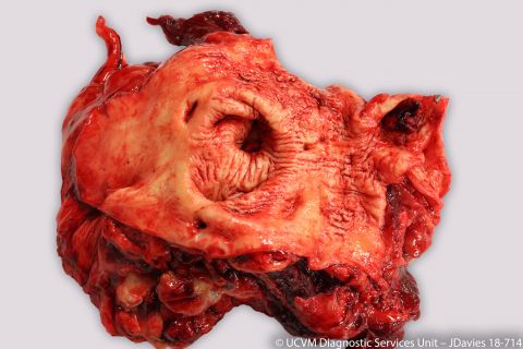
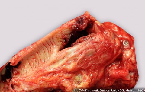
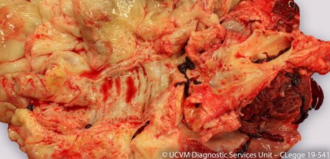
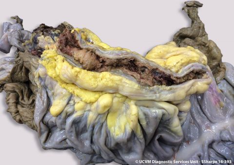
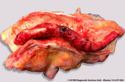
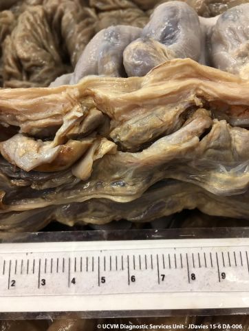
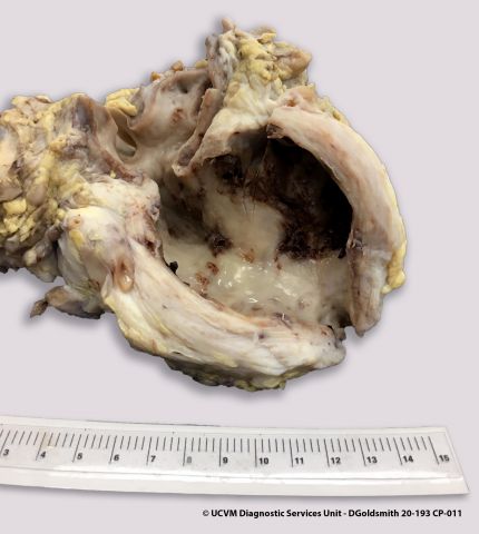
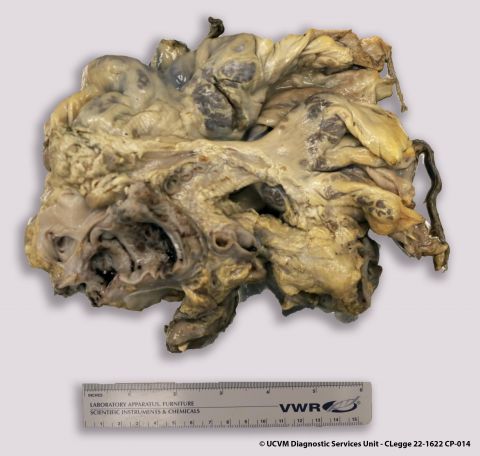
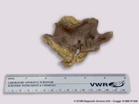
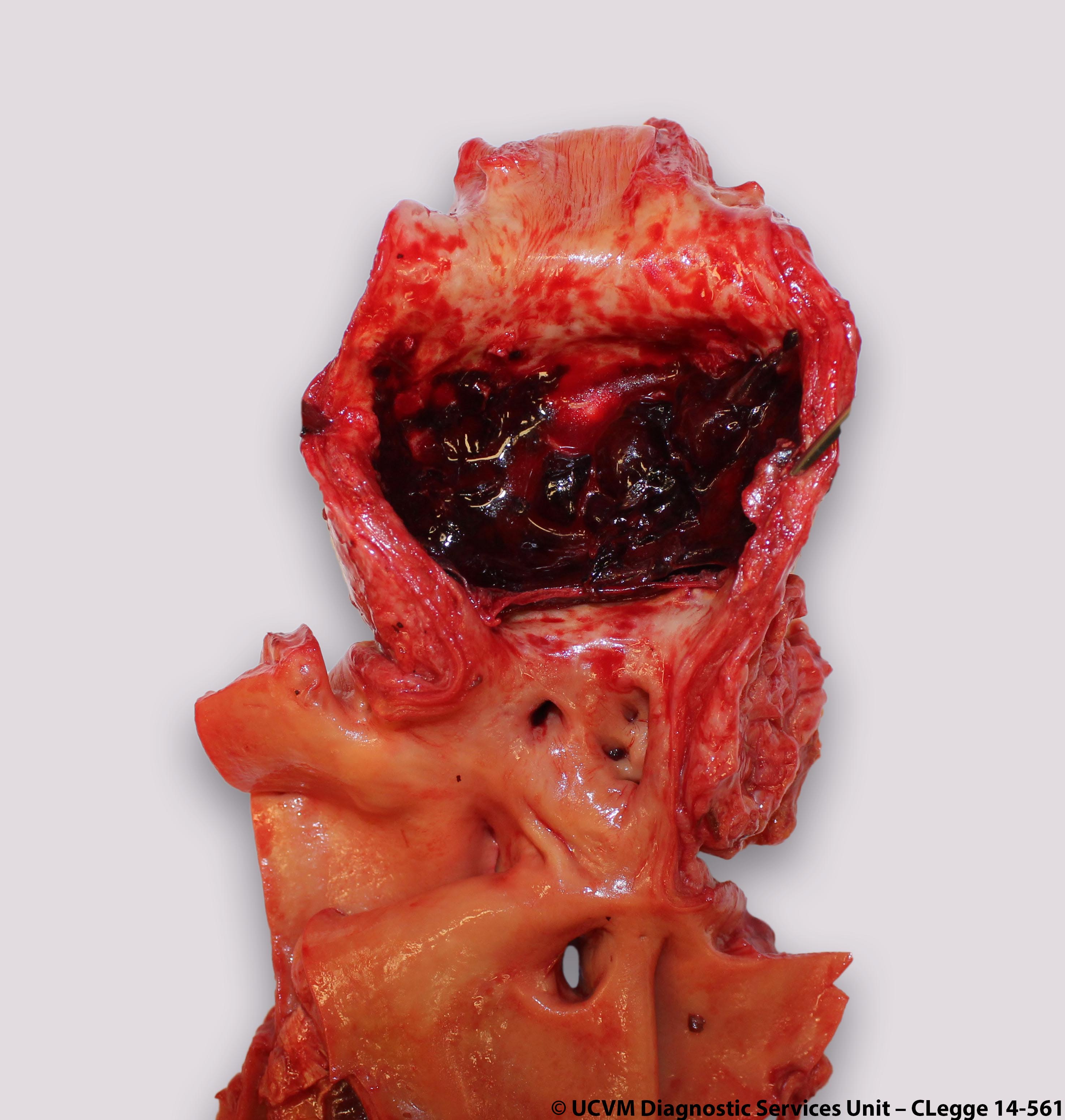
Cranial mesenteric artery
Shown:
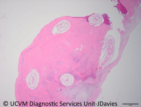
Verminous arteritis with aneurysmal dilation caused by Strongylus vulgaris
Etiology:
Strongylus vulgaris
- Up to 100% prevalence in horses
- Lesions most commonly found in the cranial mesenteric artery
- Can cause colic in horses
Lifestyle:
Horse ingests L3 larvae, which invade the small intestine, cecum and colon to molt into L4 larvae. L4 larvae penetrate arterioles to reach the cranial mesenteric artery and molt to L5 larvae after 3-4 months. L5 larvae return to the colon and cecum forming nodules, which release adults when ruptured.
Gross Lesions:
Lesions range from insignificant intimal tracks, to occlusive thrombotic lesions in the cranial mesenteric and ileocecocolic arteries
References:
Jubb, Palmer and Kennedy's Pathology of Domestic Animals